Endometriosis Mapping: What Dynamic Ultrasound Can Detect
You may have been told that endometriosis doesn't show up on imaging. That is not entirely true.
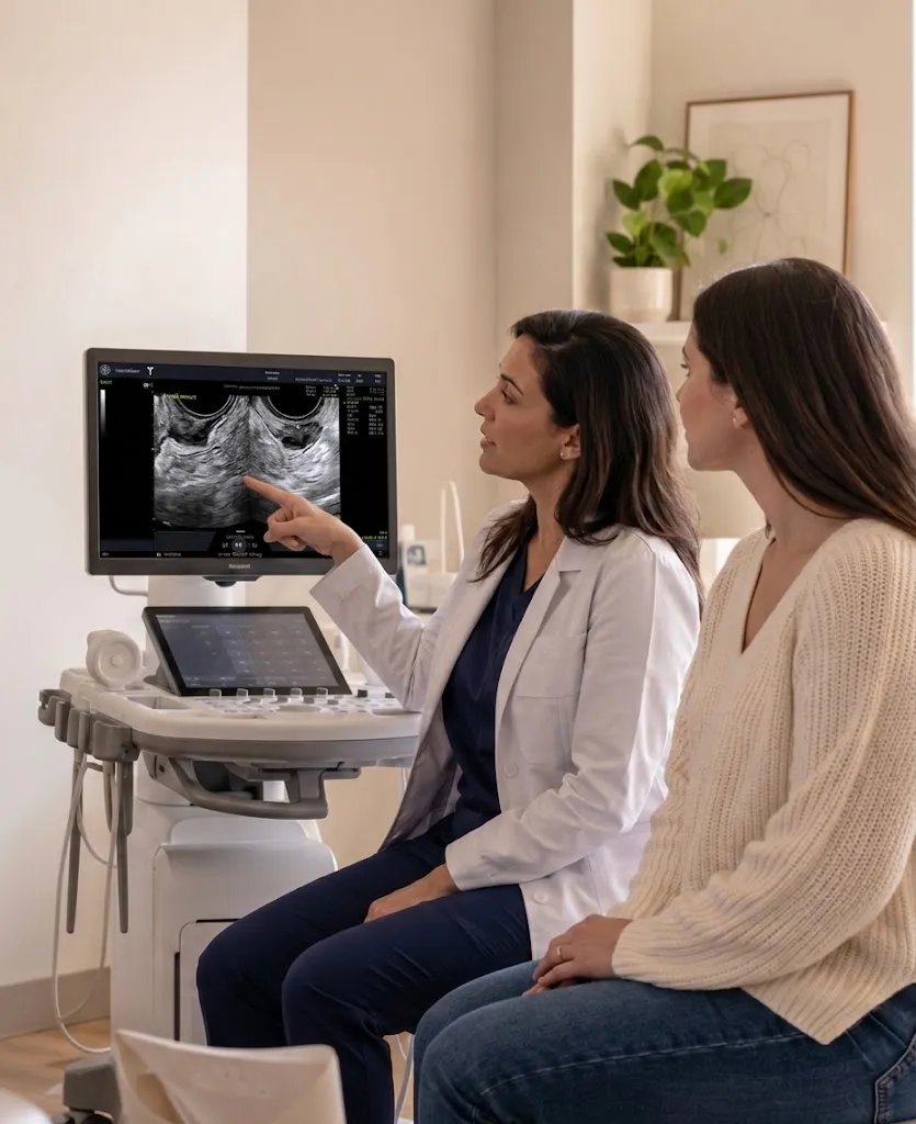
"A standard pelvic ultrasound often misses endometriosis. A dynamic ultrasound performed by a surgeon with specialized training is a different tool entirely. At WECS, this precision imaging is part of how we plan surgery — not a box we check before it."
What Dynamic Ultrasound Can Detect
Not all endometriosis appears on imaging. But for patients with deep infiltrating endometriosis, our advanced ultrasound, performed using the International Deep Endometriosis Analysis (IDEA) group protocol and Morphological Uterus Sonographic Assessment (MUSA) criteria, can reveal a great deal.
Bowel endometriosis, bladder involvement, ureteral involvement, and obliteration of the cul-de-sac (a condition called posterior cul-de-sac obliteration, caused by severe adhesions) can all be identified when the technique is right and the physician is trained to look. Research supports that advanced ultrasound is at least as effective as MRI in detecting bowel endometriosis, and in some studies, modestly superior.
What It Tells Us About Bowel Endometriosis
Beyond detecting disease, the scan can capture specific features of bowel endometriosis: the size of a lesion, its depth of penetration into the bowel wall, and its exact location. These details drive surgical decisions. They help determine whether bowel involvement will require a more complex intervention, and they allow the surgical team to prepare accordingly.
Available for Surgical Candidates
This imaging is part of the comprehensive evaluation process for WECS surgical candidates. During your evaluation, your surgeon, Dr. Victoria Vargas or Dr. Melissa McHale — both fellowship-trained minimally invasive gynecologic surgeons (MIGS) — will perform the ultrasound during your consultation. Afterward, we'll review the findings together and discuss what they mean for your surgical plan.
This is a deliberate part of how care is structured here. The surgeons at WECS have advanced training in the ultrasound technique required to map deep infiltrating endometriosis (DIE), which is distinct from general gynecologic imaging training. The technology is only as useful as the hands and eyes applying it.

Minimally Invasive Gynecologic Surgery

Minimally Invasive Gynecologic Surgery
Why It Matters for Surgical Planning
Ninety percent of surgeries at WECS involve more than one surgeon to ensure thorough removal of all lesions the first time. When bowel endometriosis is suspected or confirmed, our team includes Dr. Vincent Obias, a colorectal surgeon recognized nationally as a leading specialist in bowel endometriosis.
Knowing preoperatively what you are likely to find changes how a surgery is organized. It means the right team is in the room. It means procedures like bowel resection are discussed with the patient in advance, not decided on the table. You participate in decisions about your own surgical plan before you are under anesthesia.

Organ Preservation Is the Goal
For patients facing surgery that may involve the bowel or uterus, preservation is a priority. Dr. Obias approaches bowel endometriosis with the goal of avoiding radical procedures, including colon resection, wherever possible. When hysterectomy is considered, the surgical team weighs every option with the patient's long-term health and reproductive goals in mind.
What This Means for You
If you've been told your imaging was normal, that doesn't rule out endometriosis. It may mean the imaging wasn't performed with the right technique, by the right provider, looking for the right things.
Precision endo mapping via ultrasound at WECS is a surgical planning tool, not a screening tool. It is available to candidates who are already in the evaluation process. It is one part of our multidisciplinary approach designed to ensure that when you reach the operating room, nothing is a surprise.
.webp)